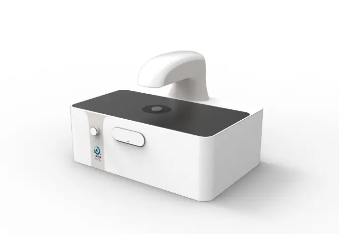
Omni Cell Imager® (OCI)
Unlock the Full Potential of Cell Imaging
It’s time to make a change
Imaging across modalities is critical—but switching between BF, DF, PC, and DIC often wastes time and introduces errors. And when new, advanced imaging modes not previously available (e.g., fused and quantitative phase imaging) are added——these challenges multiply. Cell imaging should be flexible, precise, and intuitive—but too often, it’s not. Researchers are forced to manually switch modes, swap components, realign samples, recalibrate systems, and lose valuable time in the process. The Omni Cell Imager is built to change that.
Redefine what's possible in multimodal cell imaging
Omni Cell Imager (OCI): a cutting-edge, standalone, software-defined system designed to eliminate both the technical obstacles and day-to-day hassles of cell imaging. Powered by TLI's proprietary Programoptics paradigm and advanced spatial light modulators (SLMs) algorithms, OCI meets the diverse demands of cellular and bio-tissue research. Its unmatched versatility, precision, and performance bring seamless automation and AI connection withing a single workflow.
Performance Showcase
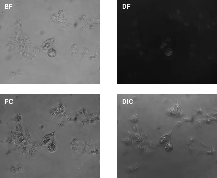
Prostate Cancer Images (BF, DF, PC, DIC)(40x)
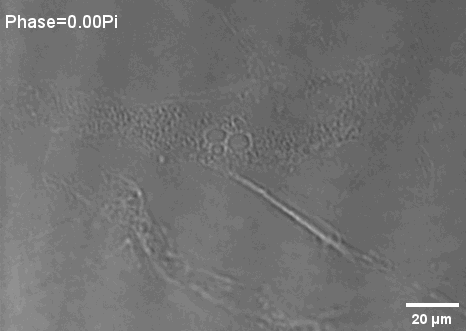
ADSC Phase Scan PC Images
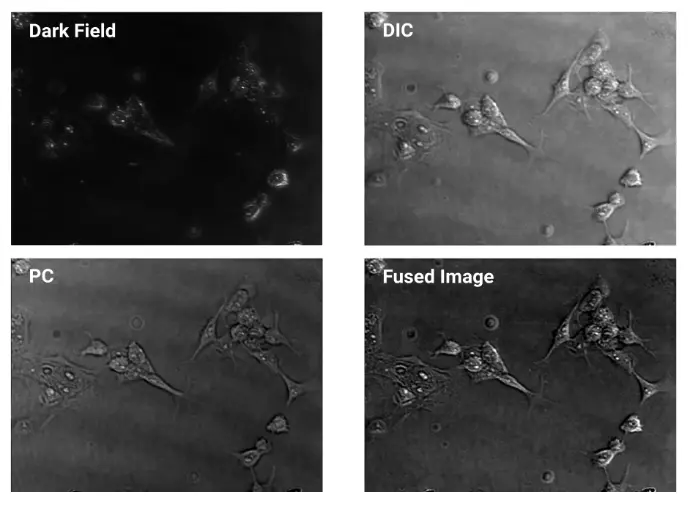
Prostate Cancer Images (DF, PC, DIC, Fused Image)
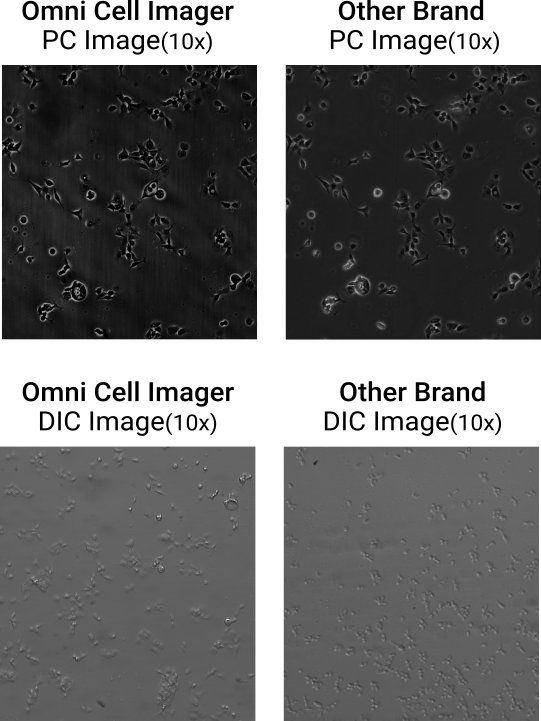
One System. Both Phase Contrast and DIC. Image Quality You Can Trust
Achieve high-contrast PC and DIC imaging on a single, software-defined microscope — with clarity rivaling traditional optical systems.
Quantitative Phase Image (QPI) with BF, DF, PC, and DIC(40x)
~0.25s to take one QPI image, ~0.6s to take all images
ADSC Images
(BF, DF, PC, DIC)
ADSC Phase Scan PC Images
Prostate Cancer Images
(DF, PC, DIC, Fused Image)

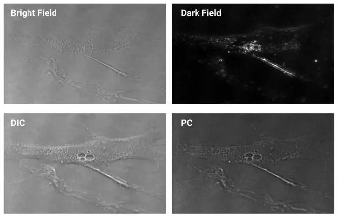
Real-Time Recording of Exosome Brownian Motion
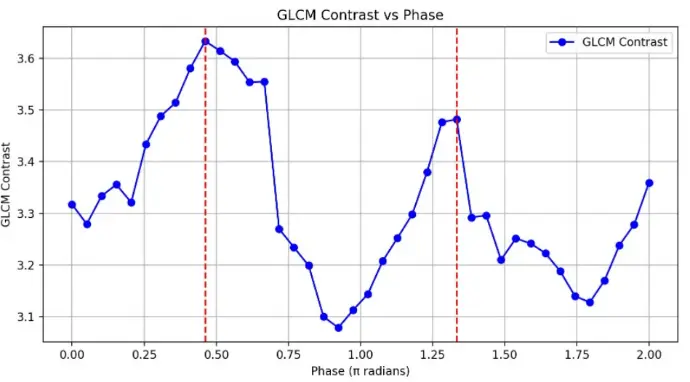
The Relationship Between GLCM Contrast and Phase
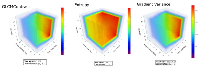
Optimal Proportion Analysis of Fused Images

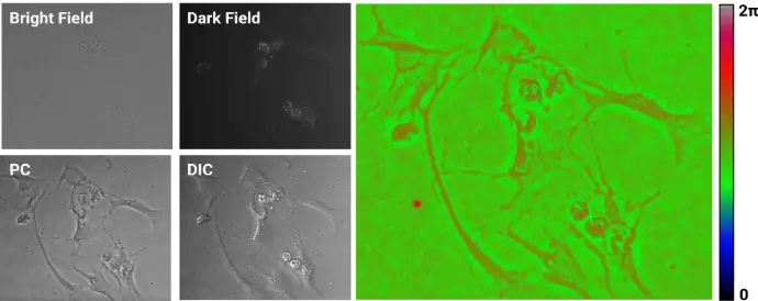
Software UI and Simultaneous Displaying 4 Modes(BF, DF, PC, DIC) of Images of The Same ROI

No More Trade-offs in Cell Imaging

Color-coded max projection from a 0.1π phase-scan stack, capturing subtle refractive-index variations in a pine stem tissue slice

Phase Scan Phase Contrast (PSPC) Image
Real-Time Recording of Exosome Brownian Motion
The Relationship Between GLCM Contrast and Phase
Optimal Proportion Analysis of Fused Images
Software UI and Simultaneous Displaying
4 Modes of Images of
The Same ROI

No More Trade-offs in Cell Imaging
Color-coded max projection from a 0.1π phase-scan stack, capturing subtle refractive-index variations in a pine stem tissue slice