AutoMorph Cell Imager®(AMCI)
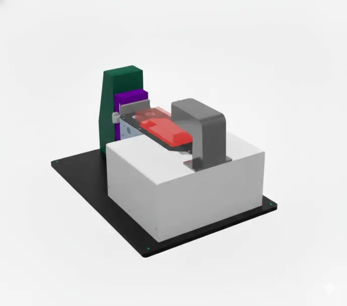
Redefining Automated Cell Culture Monitoring
In the rapidly advancing field of cell therapy, ensuring consistent and precise monitoring of cell cultures is critical. Traditional optical microscopy often struggles to meet the demands of automated environments due to mechanical switching, sample repositioning, and limited multimodal capabilities. The AutoMorph Cell Imager, powered by the innovative Programoptics paradigm and leveraging advanced spatial light modulators (SLMs), addresses these challenges by delivering a revolutionary solution for cell culture monitoring in automated production lines.
Performance Showcase
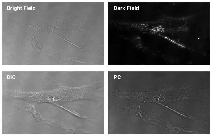
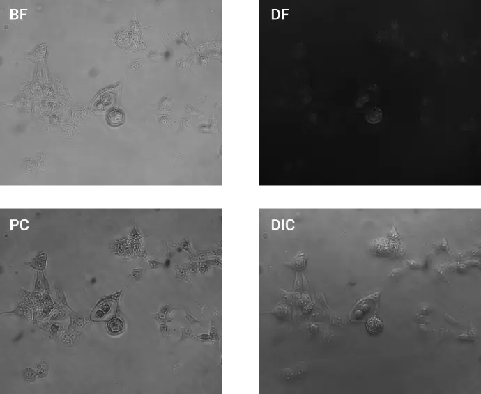
ADSC Images
(BF, DF, PC, DIC)
ADSC Phase Scan PC Images
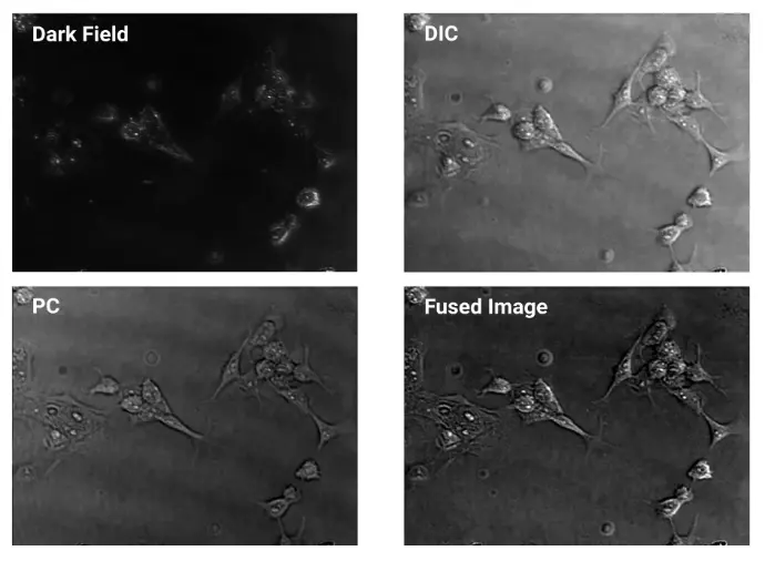
Prostate Cancer Images
(DF, PC, DIC, Fused Image)
Prostate Cancer Images (BF, DF, PC, DIC)(40x)
ADSC Phase Scan PC Images
Prostate Cancer Images
(DF, PC, DIC, Fused Image)
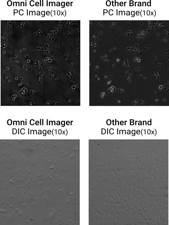
One System. Both Phase Contrast and DIC. Image Quality You Can Trust
Achieve high-contrast PC and DIC imaging on a single, software-defined microscope — with clarity rivaling traditional optical systems.

Real-Time Recording of Exosome Brownian Motion
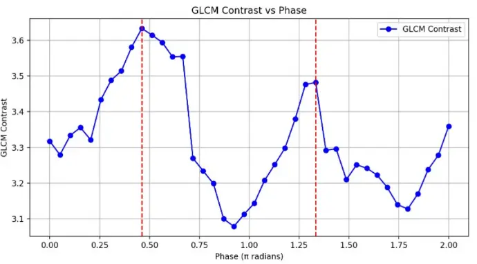
The Relationship Between GLCM Contrast and Phase
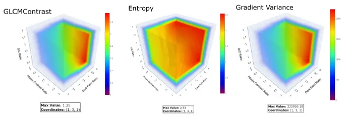
Optimal Proportion Analysis of Fused Images

Software UI and Simultaneous Displaying 4 Modes(BF, DF, PC, DIC) of Images of The Same ROI

No More Trade-offs in Cell Imaging

Color-coded max projection from a 0.1π phase-scan stack, capturing subtle refractive-index variations in a pine stem tissue slice

Phase Scan Phase Contrast (PSPC) Image
Real-Time Recording of Exosome Brownian Motion
The Relationship Between GLCM Contrast and Phase
Optimal Proportion Analysis of Fused Images
Software UI and Simultaneous Displaying
4 Modes(BF, DF, PC, DIC) of Images
of The Same ROI
No More Trade-offs in Cell Imaging
Color-coded max projection from a 0.1π phase-scan stack, capturing subtle refractive-index variations in a pine stem tissue slice